Retina

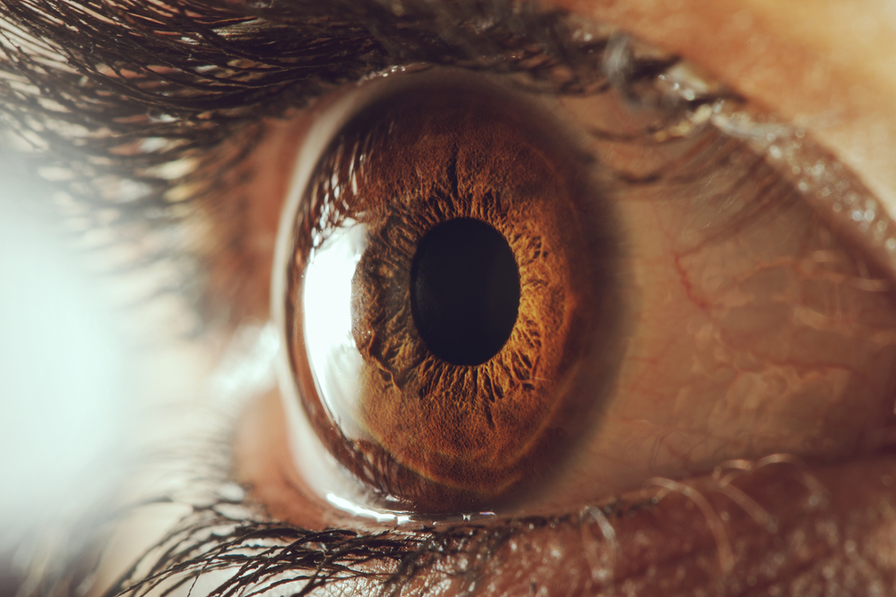
close up picture of a brown eye showing the retina.

Understanding the Retina

The retina is a fundamental structure of the eye that’s responsible for visual stability. Damage to the retina can result in a series of problems. Patients with diabetes need to be extra cautious regarding retinal eye problems. Moreover, they should seek regular eye exams to monitor the impact of this disease. If you need immediate attention or would like a second opinion. The retina eye doctors at Regional Eye Associates welcome you to contact us immediately.

What is the Retina?

The retina is a significant part of the eye that helps with vision. When you see, your eyes work to convert light into signals. These are signals that the brain then interprets and turns into the images you see.

Chart Showing the Anatomy of the Eye

This takes place in the retina, which is located in the back of the eye. The retina is the sensory membrane that lines the inner surface of the back of the eye.

The retina has several layers and contains millions of light-sensitive cells. These cells are called rods and cones. Those rods and cones sense the image as different intensities of light and color.

When light hits the rods or cones, your body transmits signals along the brain’s nerve centers. In order to, interpret the information of the image in the retina. Then, the brain assembles those signals to allow us to see what is before us.

Other Important Definitions for Understanding the Retina

The macula is the central portion of the retina that’s directly opposite the lens. It is rich in cones, which are the cells that enable us to see fine detail and color. There are three classes of cones. Each is most sensitive to a different color. Such as, red, green, or blue.

At the center of the macula is a small area called the fovea. Cones are most concentrated in the fovea.

Although the fovea may seem small compared to the retina. It is a necessary part of our ability to see fine detail and color.

What is a Retina Specialist?

Regional Eye Associates is proud to offer a retina specialist among our staff of qualified physicians. A retina specialist is a medical doctor trained as an ophthalmologist. One who has not only received additional fellowship training in diseases. But also, in surgery of the vitreous and retina.

Retina Specialist

Gary J. Miller, MD, is the retina specialist at Regional Eye Associates. Dr. Miller joined Regional Eye Associates in 2011. He not only specializes in diabetic eye disease. But also, macular degeneration, retinal detachment, macular holes, and many other vitreoretinal diseases.

Dr. Miller has successfully treated patients with retinal disorders and diseases. He is also an expert on conditions of the retina. He develops a personalized treatment plan for each patient. While using his medical expertise combined with the latest technology available for treatment.

What are Retina Problems I Should Know About?

There is a wide variety of retina conditions, problems, and diseases. If you are concerned you may be experiencing any of these conditions or diseases. Make an appointment at Regional Eye Associates in Morgantown with one of our expert ophthalmologists.


Diabetic Retinopathy

Diabetes can cause many health problems. Including, diabetic retinopathy. There are two main stages of diabetic retinopathy.

The first is non-proliferative diabetic retinopathy (NPDR). And, the later stage is proliferative diabetic retinopathy (PDR). Diabetic retinopathy occurs when the blood vessels in the retina deteriorate due to high blood sugar levels.

When this happens, the blood vessels begin to swell and leak. Which, leads to significant vision loss if it’s left untreated. However, with early diagnosis and treatment, patients can avoid further vision loss


Macular Degeneration

The most common age-related eye disease is age-related macular degeneration (AMD). Which, affects an estimated 9 million Americans a year. This condition is the leading cause of vision loss in people who are 50 or older.

Macular degeneration is the deterioration or breakdown of the eye’s macula. Which, causes vision problems. When you have AMD, you lose your central vision. Meaning you can’t see fine details. But, you’ll still be able to use your peripheral vision without a problem.


Macular Edema

Macular edema occurs when there is fluid or swelling of the macula or central portion of the retina opposite the lens. It may also include fluid that collects underneath the macula. When this happens, it causes distortion and blurred vision. Macular edema has several causes. Including, diabetes. Which can occasionally occur after cataract surgery


Macular Hole

A macular hole can occur when there is a small break in the macula. The macula is located in the center of the eye’s light-sensitive tissue. Which is the retina. You can treat a macular hole with a vitrectomy.

Having a vitrectomy involves putting a gas bubble in the eye and removing the vitreous gel in the eye. This flattens the macular hole and keeps it in place while your eye heals.

The gas bubble will go away on its own. After a vitrectomy, you must remain facedown for a week or more. This is the only way to ensure the gas bubble remains in place while you’re healing.


Flashes & Floaters

For most people, flashes and floaters are a normal part of what they see. These are the spots or lines that seem to float in your field of vision.

If you start seeing unexpected flashes of light in your peripheral vision. This could be a sign of a problem with your retina. You may see them accompanied by floaters or floating spots. Or lines that show up in your field of vision.

Seeing unexpected flashes of light could not only signify conditions like retinal detachment. But also, macular degeneration or vitreous detachment. See an eye doctor as soon as possible to determine the cause.


Retinal Detachment

A retinal detachment is considered a medical emergency. It occurs when the retina pulls away from the back of the eye.

A surgeon must surgically reattach the retina as soon as possible if this happens. If left untreated, a retinal detachment can result in permanent vision loss. 

Macular Hole

A macular hole can occur when there is a small break in the macula. The macula is located in the center of the eye’s light-sensitive tissue. Which is the retina. You can treat a macular hole with a vitrectomy.

Having a vitrectomy involves putting a gas bubble in the eye. And, removing the vitreous gel in the eye. This flattens the macular hole and keeps it in place while your eye heals.

The gas bubble will go away on its own. After a vitrectomy, you must remain facedown for a week or more. This is the only way to ensure the gas bubble remains in place while you’re healing.

Macular Edema

So, macular edema occurs when there is fluid or swelling of the macula. Which is the central portion of the retina opposite the lens. It may also include fluid that collects underneath the macula.

When this happens. It causes distortion and blurred vision. Macular edema has several causes. Including diabetes. And, can occasionally occur after cataract surgery.

Wet AMD

With wet AMD, this is a less common form. But, it’s very serious. With wet AMD. Abnormal blood vessels start growing under the retina.

These blood vessels aren’t as strong as normal ones. And, they may leak blood. The leaking blood vessels can cause scarring on the macula. If you have wet AMD, you’ll lose your vision much faster than you would with dry AMD.

Treating Wet AMD

To treat wet AMD, your ophthalmologist may prescribe anti-VEGF medication. Moreover, this treatment works because it reduces the number of abnormal blood vessels on the retina. While, slowing any leaking from the new blood vessels.

One anti-VEGF treatment you may undergo is EYLEA. EYLEA. Which, is a prescription medication injected into the eye. With continued treatments, patients maintained improvement in their vision. In fact, it could be up to 4 years when they used EYLEA.

Another treatment for some types of wet AMD is laser surgery. Like photodynamic therapy. During this procedure. The surgeon shines a light beam on the abnormal blood vessels. By shining the light, you can reduce the number of blood vessels on the retina. And, slow down their leaking. With this in mind, you should talk to your ophthalmologist about what treatment option may be best for you.

Dry AMD

There are two forms of macular degeneration. They are dry and wet. The dry form is more common. Almost 80% of people with AMD have it. Dry macular degeneration occurs when parts of the macula become thinner. Which leads to the development of a protein called drusen.

Unfortunately, there is no way to treat dry AMD. But, some people may benefit from taking a specific combination of nutritional supplements. The AREDS and AREDS 2 study found that this combination may slow down dry AMD. By taking the vitamins and minerals daily. 

What is a Retinal Vein and Artery Occlusion

A retinal artery occlusion occurs when the retinal artery that carries oxygen to the retina is either entirely or partially blocked. Blocking the retinal artery means that the retina is not receiving oxygen. And, may result in severe vision loss if left untreated.

Vein occlusions can not only be caused by high blood pressure. But also, diabetes, age, and blood clotting disorders. Symptoms of a retinal artery occlusion include experiencing a sudden painless loss of vision in one eye. The part of the retina that’s affected by the blocked vessels determines how much vision loss occurs and where it is.

Retinal Vein Occlusion

Occlusions of the vessels at the point of entry to or exit from the eye are not only called central retinal vein occlusion. But also, central retinal artery occlusion.

A branch retinal vein occlusion or branch retinal artery occlusion is a blockage in the eye. That only affects part of the eye. The severity of the blockage also determines the damage.

The vision loss that patients can suffer ranges from mild to blindness. Our team at Regional Eye Associates will evaluate your condition. All by using state-of-the-art imaging techniques during an eye exam. These may include optical coherence tomography (OCT) and fluorescein angiogram (FA).

How Do You Treat Retinal Vein and Artery Occlusions?

Although there is no way to unblock retinal veins. It is possible to treat the health problems. Especially, those that caused the retinal vein occlusion. A retinal vein occlusion may cause blood vessels in the retina to leak.

These leaking blood vessels have more pressure on them because of increased blood flow. The leakage from these blood vessels may lead to macular edema.

Focal Laser Therapy

Your eye doctor may recommend laser treatments like focal laser therapy where there’s swelling. All in the hopes of reducing macular edema. They may also try anti-vascular endothelial growth factor drugs (VEGF). Which, targets VEGF. A growth factor that causes macular edema.

There are no treatment options for retinal artery occlusions. It is vital to see your eye doctor for regular appointments. Above all, they can monitor your eyes.

Proliferative Retinopathy

Monitoring your eyes will ensure that the lack of blood flow in the eye doesn’t lead to a condition called proliferative retinopathy. Proliferative retinopathy is a condition that leads to the creation of new blood vessels in the eye.

These blood vessels are quite weak and may not only bleed. But also, cause a painful increase in the eye’s amount of pressure. Treating proliferative retinopathy requires prompt surgical treatment. This may include:

Photocoagulation

Photocoagulation is also known as focal laser treatment. It’s used to slow or stop any leaking fluid or blood in the eye. During focal laser treatment, leaks in the eye use laser burns to treat abnormal blood vessels.

Panretinal photocoagulation

Panretinal photocoagulation is also known as scatter laser treatment. It works to shrink abnormal and weak blood vessels. This procedure involves treating areas of the retina that aren’t near the macula. As a matter of fact, it works with laser burns that are scattered.

Laser burns help to shrink and scar the new blood vessels. This treatment is often done in sessions of two or more at your eye doctor’s office.

Undergoing these surgical procedures can slow down or stop the progression of retinopathy. But, they are not a cure. Future vision loss and retinal damage could still occur later on.

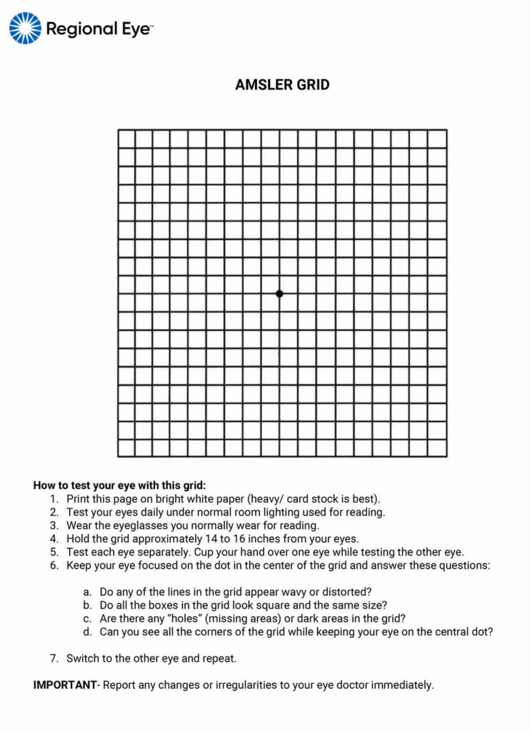
Click on the Amsler Grid below for a larger/printable version.

Amsler Grid Test


Want more information about how to treat a retinal condition? Schedule an appointment with Regional Eye Associates today.

Request Appointment Patient Portal Payment